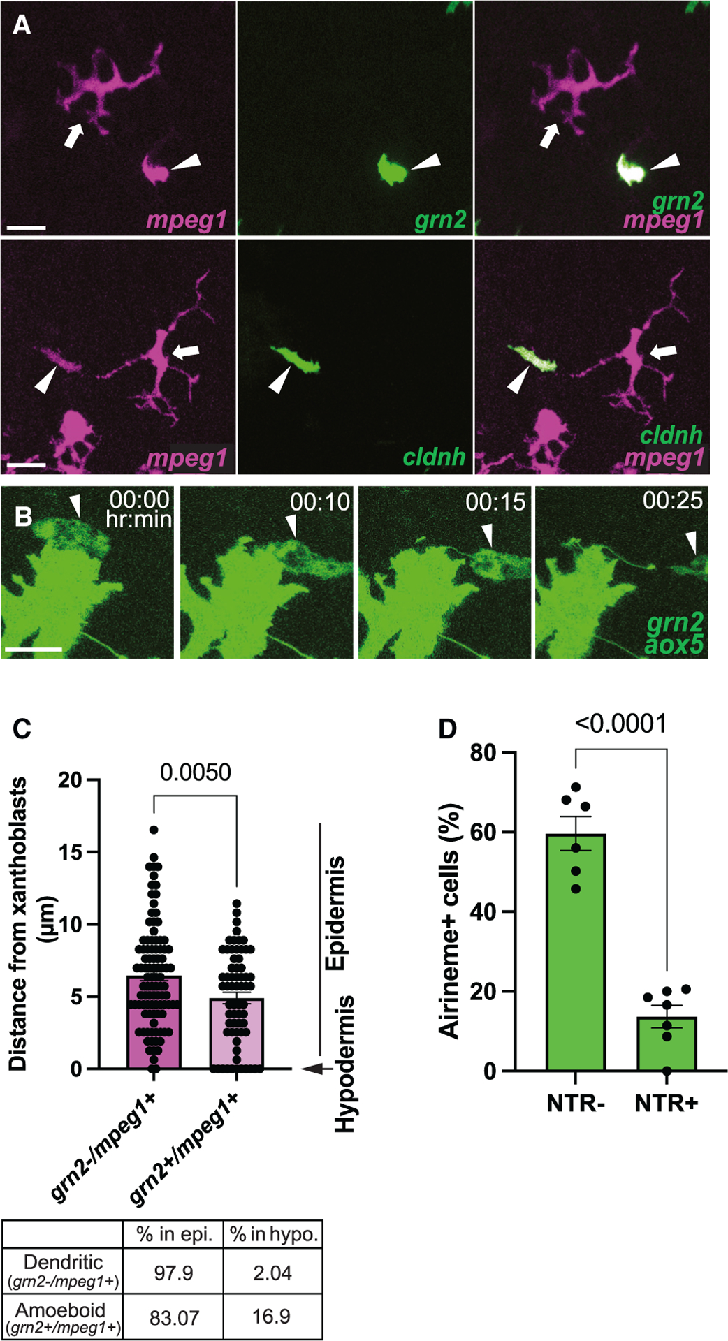
Figure 2. Airineme pulling ameboid macrophages overlap with ectoderm-derived metaphocytes.

(A) Coexpression of metaphocyte markers, grn2 and cldnh, in ameboid macrophage subpopulation (mpeg1+, arrowheads). Note that dendritic (mpeg1+, arrows) macrophages do not express metaphocyte markers.
(B) Still images from a time-lapse movie showing airinemes being pulled by grn2+ metaphocytes.
(C) Similar to the ameboid macrophage population, metaphocytes are more frequently located in the hypodermis.
(D) Airineme extension was quantified by counting the number of cells that extended airinemes at least once out of all imaged cells. Xanthoblasts of metaphocyte depleted fish (NTR+) were less likely than controls (NTR–) to extend airinemes; n = 6 (controls), n = 7 (depleted) time-lapse positions, three trunks each. The few airinemes extended in the depleted were associated with residual metaphocytes (Video S4). Statistical significances were assessed by Student t test. Scale bars, 20 μm (A), 10 μm (B). Bars display mean, and error bars display SEM.
