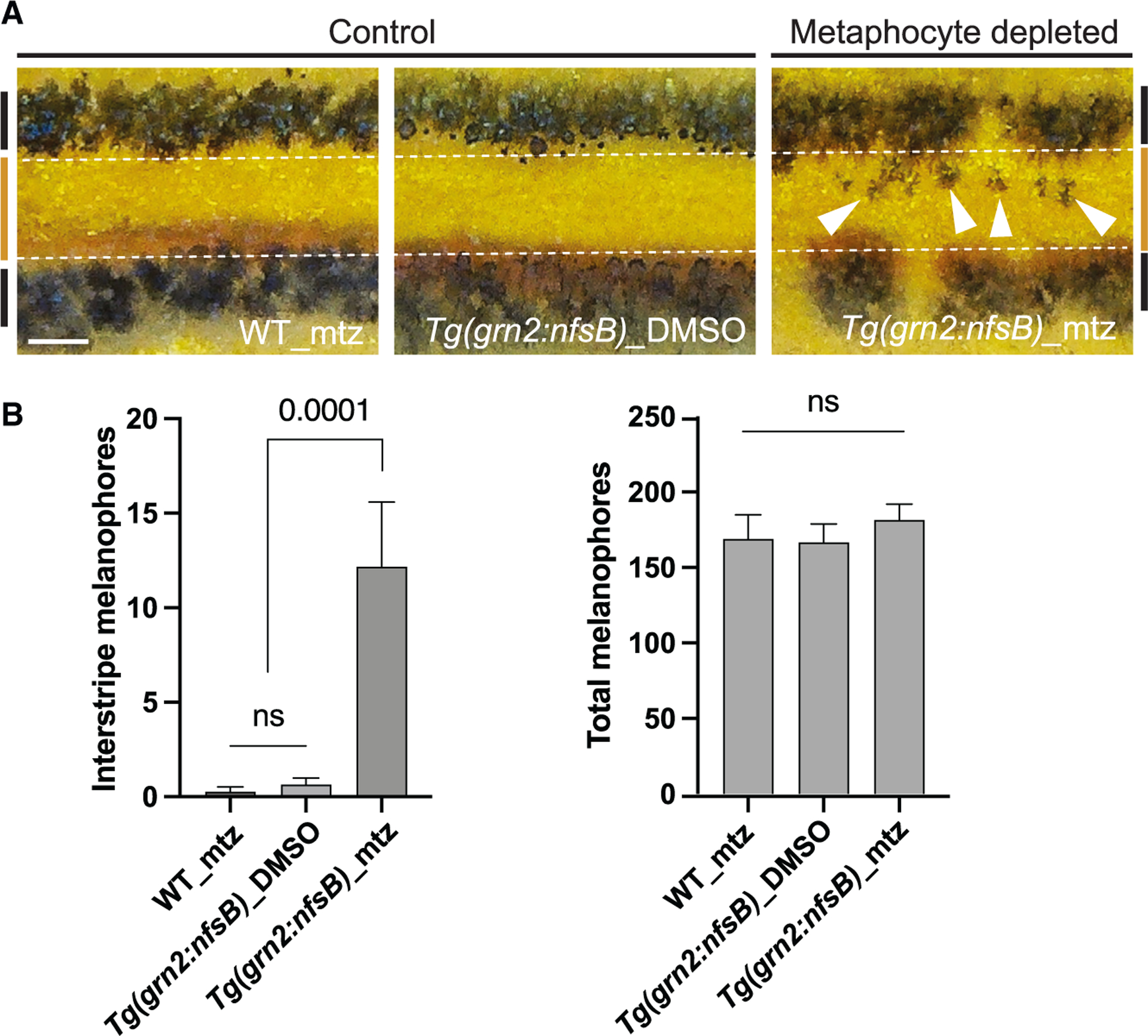
Figure 3. Metaphocyte-mediated airineme extension is responsible for melanophore pattern formation.

(A) In two controls, melanophores reside in the stripe region (denoted by black bars at far left and right, and white dotted lines) and the border of the interstripe (orange bar). Metaphocyte depletion results in melanophores retention in the interstripe (arrowheads).
(B) Numbers of melanophores in the interstripe. Metaphocyte-depleted fish (Tg(grn2:nfsB)_mtz) had significantly more melanophores in the interstripe than the controls (WT_mtz and Tg(grn2: nfsB)_DMSO) and displayed a disorganized pigment pattern, although total melanophore numbers did not differ; data are means ± SEM. At stage 12, standardized standard length (SSL); n = 4, WT_mtz, n = 15, Tg(grn2:nfsB)_DMSO, n = 11, Tg(gnr2:nfsB)_mtz trunks. Statistical significances were assessed by Student t test. Scale bar, 200μm (A). mtz, metronidazole.
