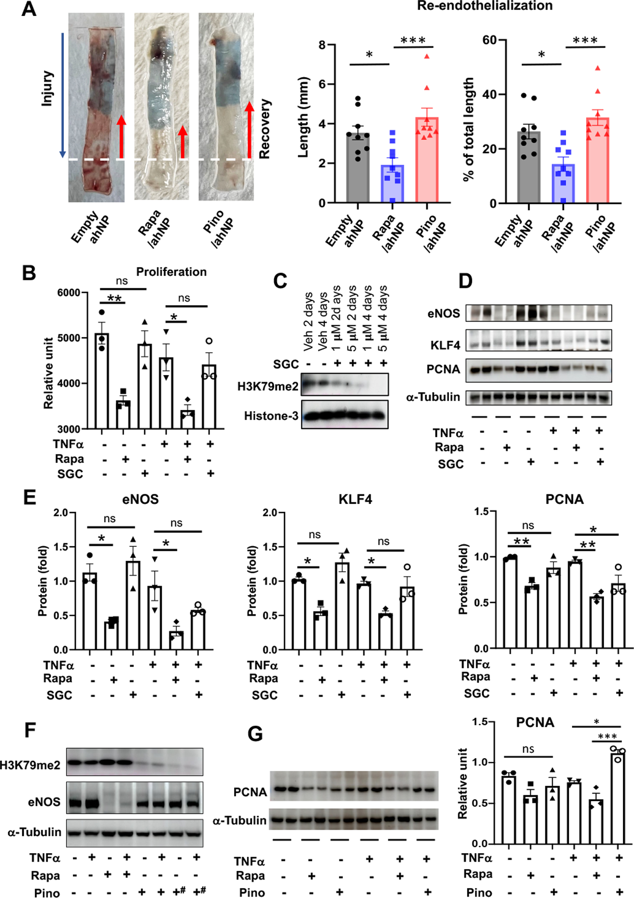
Figure 5. Pinometostat delivered by Epi^NanoPaint is not toxic to the endothelium.

A. Re-endothelialization. The endothelium was damaged as far as the dotted white line in the IH model of rat common carotid artery injury and grew back (after 7 days) as much as the length of red arrow marking the unstained segment. The remainder damaged segment was stained blue. Drug dose: 0.3 mg/kg pinometostat; 0.018 mg/kg rapamycin. Mean ±SEM, n= 8 or 9 Sprague-Dawley rats. One-way ANOVA and Tukey test: *p<0.05, ***P<0.001.
B-E. In vitro assays using the DOT1L-selective inhibitor SGC0946. Human umbilical vein endothelial cells were cultured in Lonza EGM-2 growth medium until ~80% confluency and starved in basal medium overnight. Rapamycin (final 0.5 μM) or SGC0946 (final 5 μM) or vehicle (veh, equal volume of DMSO) was added to the starved cells and incubated for 2 days (note different concentrations of SGC and pretreatment days tested in C). The cells were then treated with TNFα (20 ng/ml) for 4h followed by Cell Titer Glo viability assay and immunoblot assay. In D, duplicate immunoblot bands represent two separate experiments. Quantification: Mean ± SEM, n = 3 independent repeat experiments; oneway ANOVA and Tukey test: *p<0.05, **P<0.01.
F-G. In vitro assays using the DOT1L-selective inhibitor Pino. Human umbilical vein endothelial cells were cultured in Lonza EGM-2 growth medium until ~80% confluency and starved in basal medium overnight. Rapamycin (final 0.5 μM) or vehicle (equal volume of DMSO) was added to the starved cells and incubated for 2 days. Pretreatment with Pino (final 0.5 μM) continued for 3 or 5 days (hashtags in F). The cells were then treated with TNFα (20 ng/ml) for 4h followed by immunoblot assay. In G, cells were pretreated with 0.5 μM Pino for 5 days and 0.5 μM Rapa for 2 days. Duplicate bands on the immunoblot represent two separate experiments. Quantification: Mean ± SEM, n= 3 independent repeat experiments; one-way ANOVA and Tukey test: *p<0.05, ***P<0.001.
