
Figure 4: NET1A-CASK interaction competes with NET1A-importin binding and is regulated in an opposite manner by NET1 mRNA location.
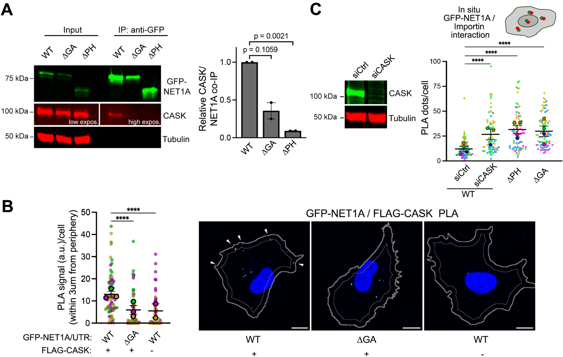
(A) Representative Western blot and quantification of relative CASK binding to GFP-NET1A from co-immunoprecipitation experiments of the indicated MDA-MB-231 cell lines. n=2. (B) In situ detection of interaction between GFP-NET1A and FLAG-CASK, by PLA in the indicated cell lines. Cyan dots: PLA signal; blue: DAPI; thick gray outline: cell boundary; thin gray outline: inner boundary 3μm from periphery. n=45–72 in 3 independent experiments. (C) Western blot showing efficiency of CASK knockdown upon siRNA treatment and quantification of in situ interaction between GFP-NET1A and FLAG-importin β1, by PLA of the indicated cell lines (see Figure 3C). n=78–92 in 4 independent experiments. In superplots, data points from individual replicates are color coded, and large, outlined color dots indicate the mean of each replicate. Error bars: SEM. p-values:, ****<0.0001 by one-way ANOVA (A, C) or Kruskal-Wallis test (B). Scale bars: 10μm.