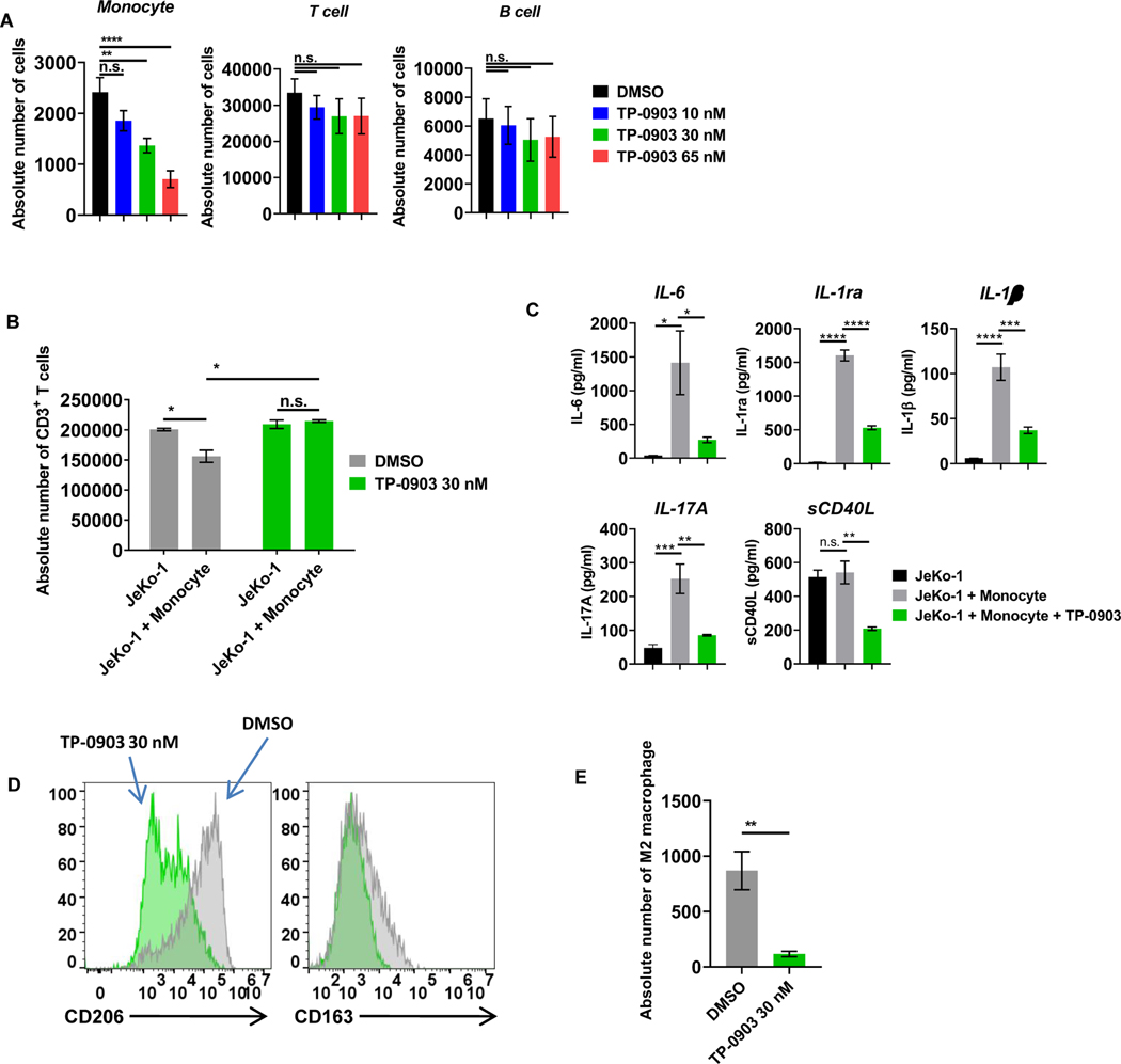
Figure 5: AXL inhibition with TP-0903 preferentially targets monocytes and overcomes monocyte-induced CART-cell suppression.
A, Peripheral blood mononuclear cells derived from healthy donors were treated with increasing doses of TP-0903 (10–65 nM) for 24 hours, and the absolute number of cells was assessed by CountBright bead quantification with flow cytometry (n.s. not significant, ** p<0.01, **** p<0.001, one-way ANOVA, n=3 biological replicates). B, CART19 cells, JeKo-1 cells, and monocytes were co-cultured for 5 days and analyzed for the absolute number of CD3+ T cells by CountBright bead quantification (* p<0.05, t-test, n=3 biological replicates). C, Supernatants that were harvested from experiment in Fig. 5B were analyzed with 38-mulitplex. (* p<0.05, ** p<0.01, *** p<0.001, **** p<0.0001, one-way ANOVA, n=3 biological replicates). D, CD14+ cells were stained for M2-related markers CD206 and CD163 and analyzed via flow cytometry. E, CART19 cells, JeKo-1 cells, and M2-like macrophages were co-cultured for 5 days and analyzed for the absolute number of CD14+ cells by flow cytometry (** p<0.01, t-test, n=3 biological replicates). Data are plotted as means ± SEM.

