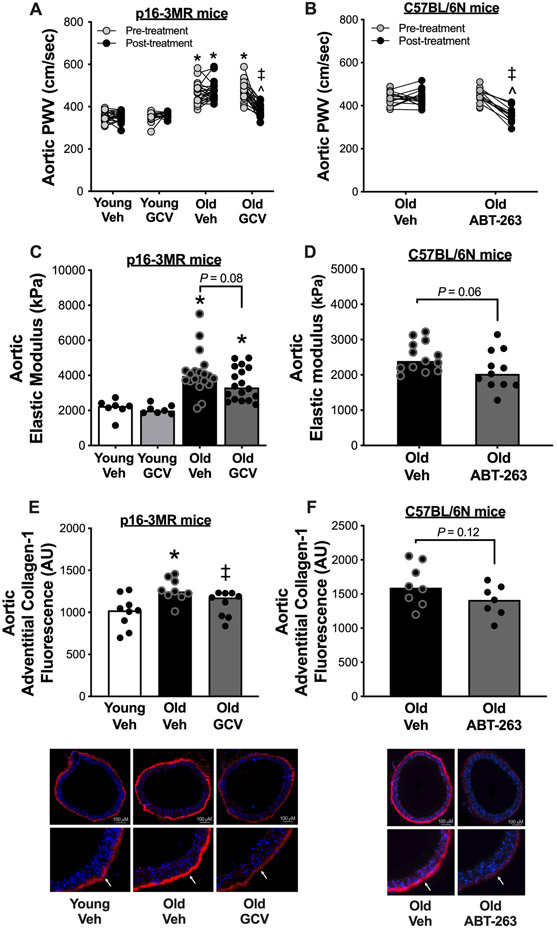
Figure 1. Cellular senescence mediates aortic stiffening with aging and senolytic treatment lowers aortic stiffness in advanced age.
In young (6-8 months) and old (27-29 months) male and female (sexes combined) p16-3MR mice treated with vehicle (Veh; sterile saline) or ganciclovir (GCV, in sterile saline) at a dose of 25 mg/kg/day for five consecutive days via intraperitoneal injection: (A) Aortic pulse wave velocity (PWV) before and after treatment. (C) Aortic elastic modulus at time of sacrifice. (E) Immunofluorescence staining for Type-1 Collagen in the adventitial layer of aortic rings collected at time of sacrifice. In old male C57BL/6N mice treated with vehicle (10% ethanol; 30% PEG400; 60% Phosal 50PG) or ABT-263 (in the vehicle) at a dose of 50 mg/kg/day via oral gavage following a one week on – two weeks off – one week on dosing regimen: (B) Aortic pulse wave velocity before and after treatment. (D) Aortic intrinsic mechanical wall stiffness at time of sacrifice. (F) Immunofluorescence staining for type-1 collagen in the adventitial layer of aortic rings collected at time of sacrifice. All data are mean ± SEM. N = 16-23/group, p16-3MR study aortic PWV. N = 15-20/group, p16-3MR study aortic intrinsic mechanical wall stiffness. N = 10/group, p16-3MR study aortic adventitial type-1 collagen fluorescence. N = 14-16/group, ABT-263 study aortic PWV. N = 11-14/group, ABT-263 study aortic intrinsic mechanical wall stiffness. N = 9/group, ABT-263 study aortic adventitial type-1 collagen fluorescence. * P < 0.05, effect of aging within group; ‡ P < 0.05, effect of treatment within age group; ^ P < 0.05, effect of time.

