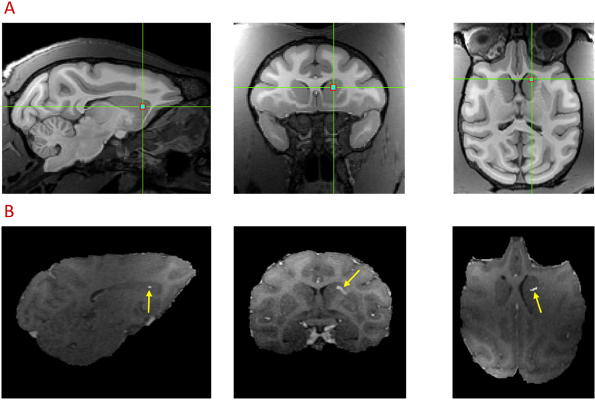
Fig. 2.

FUS target planning (A) and confirmation (B) using Gadolinium enhanced MRI images in sagittal, coronal and axial planes. The green-crossed point indicates the pre-treatment target of FUS exposure using Brainsight Navigation System (A). The hyper-intense voxels (yellow arrow) denote the region of BBB opening (B). Subtraction between Gadolinium enhanced T1 images collected at post FUS + MB and baseline was performed, and the difference in target area was shown with an overlay on post Gadolinium T1 images after FUS-induced BBB opening.
