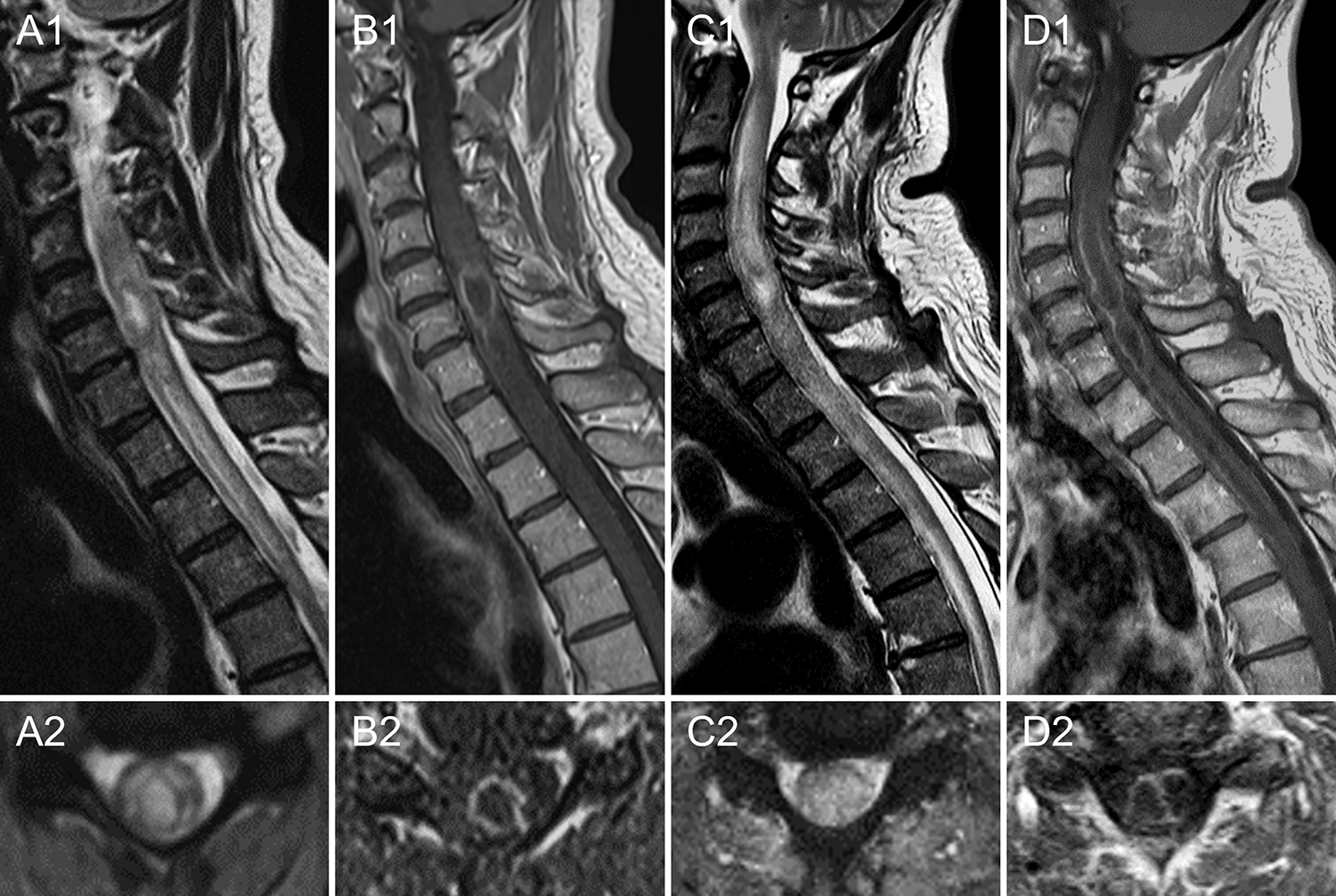
Figure 3 -. Intramedullary spinal cord abscess misdiagnosed as seronegative NMOSD.

A 54-year-old Caucasian man developed severe neck and shoulder pain, followed by progressive weakness of the left limbs and sensory loss in both hands over 3 days. His medical history was unremarkable, except for several minor dentistry procedures over the prior months. Spinal cord MRI showed a longitudinally extensive T2-hyperintense lesion from C3 to C7 with a central core of higher T2-hyperintensity (A) surrounded by an irregular ring of enhancement on post-gadolinium T1 sequences (B). CSF examination revealed 85 white blood cells and protein elevation (90 g/dl). Extensive infectious screening on both serum and CSF (including blood and CSF culture) was unremarkable. Testing for aquaporin-4-IgG and myelin oligodendrocyte glycoprotein-IgG was negative on serum and CSF by live cell-based assay. He was started on intravenous methylprednisolone (1 g daily for 5 days) with no improvement. A new MRI showed extension of the lesion to the cervicomedullary junction and the thoracic spinal cord to the T4 level (C). A longitudinal extension of the ring enhancement was also observed after gadolinium administration, which appeared bilobate on axial images at cervical level (D). A working diagnosis of seronegative NMOSD was made and plasma exchange was started. Given further clinical deterioration after two plasma exchange sessions (the patient became tetraplegic), a spinal cord biopsy was performed. Cultures from intralesional material came back positive for Haemophilus Influenzae, and antibiotic therapy was started with slow improvement. No fever or increased serum procalcitonin levels were observed before the spinal cord biopsy.
