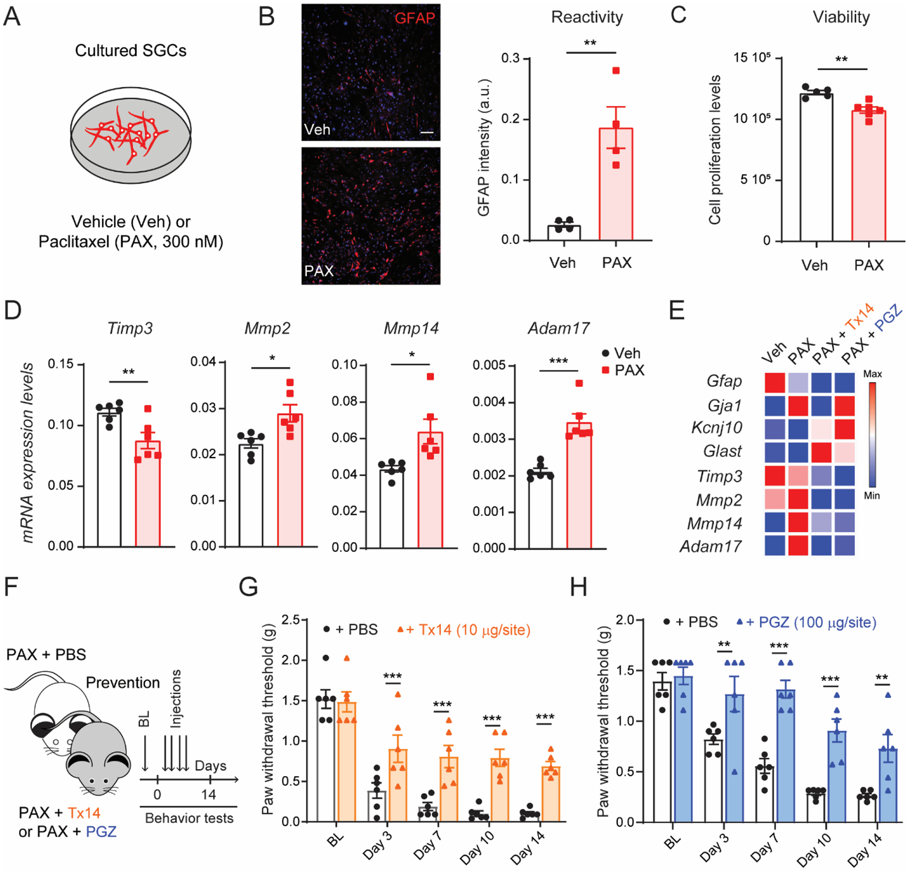
Fig. 6. Transcriptional analyses of TIMP3 signaling in cultured SGCs after paclitaxel treatment.

(A) Schematic of the experimental design used in cultured SGCs. (B) Representative image and quantification of immunofluorescence intensity of GFAP protein in SGC culture after 24 h of incubation with paclitaxel (PAX, 300 nM) or vehicle control (Veh; n=4). (C) Quantification of SGC culture viability 24 h after PAX or Veh treatment (n=6). (D) Quantification of mRNA expression levels of Timp3, Mmp2, Mmp14 and Adam17 in SGC culture after PAX or Veh incubation (n=6). (E) Heat map of mRNA expression of SGC and metalloprotease signaling markers in SGC culture after incubation with prosaptide Tx14 (1 μM) or pioglitazone (PGZ, 10 μM) and PAX compared to vehicle (n = 3). (F) Schematic illustrating the timeline of Tx14, PGZ, or PBS concomitantly treated with PAX, and the behavioral assay. Repeated injections of (G) Tx14 (10μg/site, i.t.) or (H) PGZ (100μg/site, i.t.) prevent paclitaxel-induced mechanical allodynia (n=6). BL = baseline. Data are expressed as mean ± SEM and statistically analyzed by two-tailed t-test (B, C, D), and Two-way ANOVA followed by Sidak’s post hoc test (G, H): *P < 0.05, **P < 0.01, ***P < 0.001.
