Figure 1.
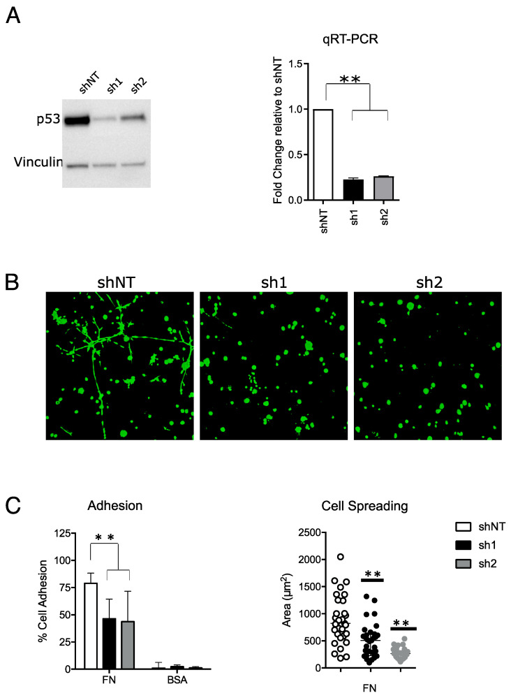
Mutant p53 confers pro-invasive phenotype in MDA-MB-231 breast cancer cell line. (A) Western blot (left) and qRT-PCR (right) of p53 in MDA-MB-231 shNT (shRNA not targeting), sh1 and sh2 at 9–10 days after shRNA transduction. Full-length blot is shown in Supplementary Figure S1A. (B) Representative 10× images of 3D Matrigel colony assay obtained with fluorescence confocal microscopy after calcein staining. Images were acquired 10 days following cell plating. (C) Left—histogram showing the percentage of cells that adhered to fibronectin (FN)-coated or control bovine serum albumin (BSA)-coated surface. Right—scatter plot showing the area of each single cell that adhered to FN-coated surface. Results are the mean of three independent biological replicates (** p-value ≤ 0.01).

