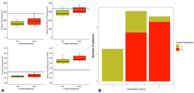
Figure 3.
(A) Comparison of quantitative OCT imaging with normative data, showing an increase in thickness and volume of the fovea, inner ring thickness (IRT) and inner ring volume (IRV) in patients with FH, with no statistically significant differences between the two groups. (B) Retinal lamination showing a higher prevalence of worse retinal lamination (groups 2 and 3) in the FH group than those without FH.

