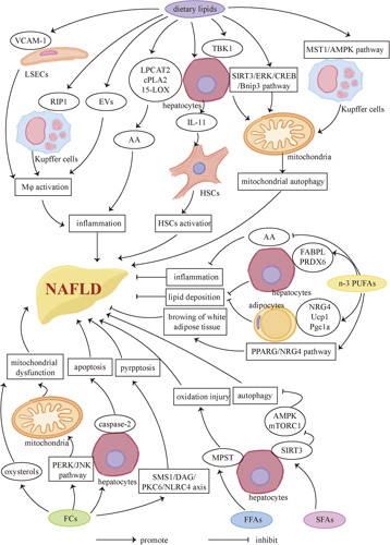
FIGURE 2.

Lipids and NAFLD. Dietary lipids have a dualistic influence on NAFLD. On one hand, excess lipids, including FCs and FFAs and so forth, especially SFAs, promote the development of NAFLD. Inflammation can be promoted by activating KCs, liver macrophages (Mφ), and stimulating the release of the proinflammatory factor AA and IL-11. Mitochondrial autophagy is also associated with one of the promoting factors of NAFLD through the macrophage stimulating 1/AMPK pathway. Excess FCs increase the occurrence of mitochondrial dysfunction and cause caspase-2–mediated hepatocyte apoptosis and SMS1/DAG/PKC6/NLRC4 axis–mediated pyroptsis. Besides, inhibition of autophagy in hepatocytes may also be one of the factors that promote NAFLD. On the other hand, increasing dietary intake of n-3 PUFAs can alleviate NAFLD mainly by inhibiting proinflammatory factors and AA, and reducing lipid deposition. Besides, n-3 PUFAs contribute to browning of white adipose tissue. Abbreviations: AA, arachidonic acid; AMPK, AMP-activated protein kinase; EV, extracellular vesicle; FC, free cholesterol; FFA, free fatty acid; JNK, c-Jun N-terminal; LOX, lipoxygenase; LPCAT2, lysophosphatidylcholine acyltransferase 2; MPST, mercaptopyruvate sulfurtransferase; mTORC1, mammalian target of rapamycin complex 1; NRG4, neuregulin 4; PERK, PKR-like endoplasmic reticulum kinase; Pgc1α, peroxisome proliferators–-activated receptor γ coactivator lα; PPARG, peroxisome proliferator– activated receptor gamma; RIP1, receptor-interacting protein 1; SIRT3, sirtuin 3; SMS1, sphingomyelin synthaseUcp1, uncoupling protein 1; VCAM-1, vascular cell adhesion molecule 1.
