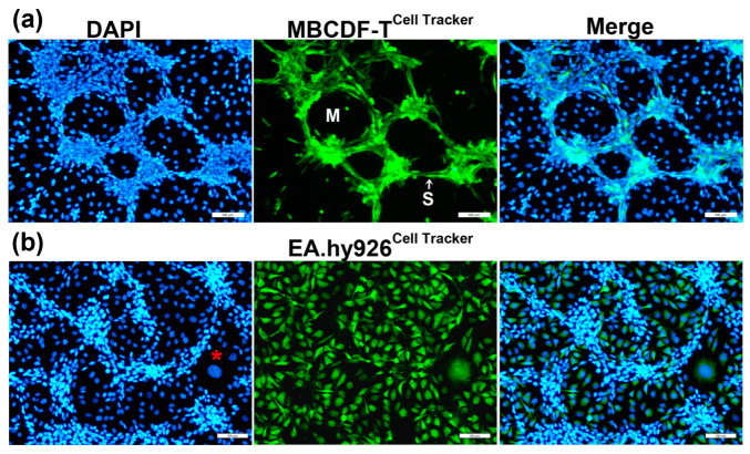
Figure 2.
TNBC cells form the tubular structures in endothelial–breast cancer co-cultures. ECs and TNBC cells co-cultures resulted in VM formation rather than endothelial angiogenesis. (a) MBCDF-T cells were labeled with a green cell tracker and then co-cultured with unlabeled ECs (EA.hy926) for 48 h. As depicted, exclusively TNBC cells were involved in the tubular-like network, displaying a spindle-like shape and conforming segments (S) and meshes (M). (b) EA.hy926 cells were labeled with the green cell tracker before co-culturing them with unlabeled MBCDF-T cells. The nuclei were counterstained with DAPI (blue channel). Giant, flattened ECs characterized by a large nucleus may be found in the co-cultures (red asterisk). Representative images were obtained via epifluorescence microscopy. Magnifications from 10× pictures are shown. Scale bar = 100 μm.

