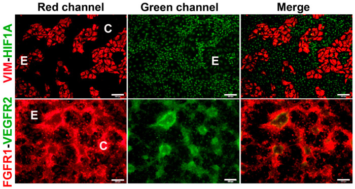
Figure 4.
Co-cultured TNBC cells and endothelial cells differentially express markers of VM and endothelium. HCC1806 cells and EA.hy926 cells were co-cultured for 48 h. Then, co-cultures were fixed and further processed for immunocytochemistry. Each slide was incubated with two primary antibodies, one made in mice and one in rabbits. Incubations were undertaken overnight at 4 °C. Mice-made primary antibodies were detected using a secondary goat anti-mouse-Cy3 antibody (red channel, left panels). Rabbit antibodies were detected with a goat anti-rabbit-FITC (green channel, middle panels). The right panels show merged pictures. As a guide, the letter “C” depicts the location of cancer cells in the segments of VM structures, while the letter “E” depicts the endothelial cells position. Cell images were captured using a conventional fluorescence microscope. Representative 10× images are shown. Scale bar = 100 μm. Vimentin—VIM; hypoxia-inducible factor-1α—HIF1; vascular endothelial growth factor receptor 2—VEGFR2; fibroblast growth factor receptor 1—FGFR1.

