Figure 5.

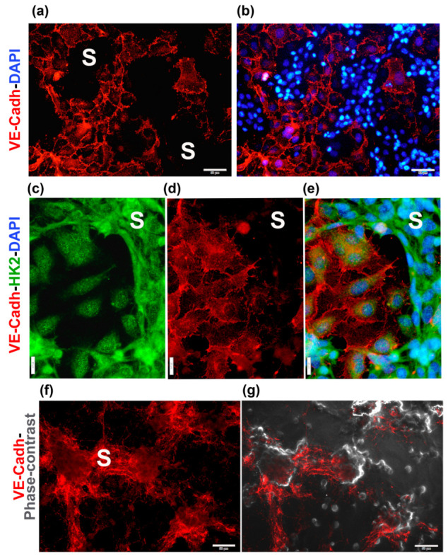
VE-cadherin and HK2 are differentially localized in the endothelial and cancer components of the co-cultures. HCC1806 cells and EA.hy926 cells were co-cultured for 48 h. Then, co-cultures were fixed and further processed for immunocytochemistry. Mouse anti-VE-cadherin (VE-Cadh) and rabbit anti-hexokinase 2 (HK2) were used as primary antibodies. Secondary antibodies were goat anti-mouse-Cy3 (red channel) and goat anti-rabbit-FITC (green channel). Junctional VE-Cadh is seen in the endothelial component of the co-cultures, while it is scarcely expressed in TNBCs (a,b,d,e). In certain portions of the preparations, nuclear and membrane localization of VE-Cadh in TNBC cells forming the vasculogenic mimicry segments (S) and branch intersections can also be clearly seen (f,g). DAPI blue staining is seen in (b,e). Cell images were captured using a conventional fluorescence microscope (a–f), while (g) is the merged image of (f) and the corresponding phase contrast photograph. Magnification is as follows: (a,b,f,g) use 20× magnification (scale bar = 50 μm); (c–e) are 40× images (scale bar = 20 μm).
