Figure 6.

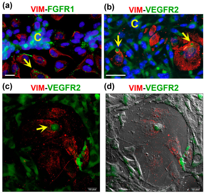
FGFR1 and VEGFR2 are localized in vesicles and vacuoles of endothelial cells in CCs. MBCDF-T and EA.hy926 cells were co-cultured for 48 h, followed by fixation and immunocytochemistry processing. (a) The primary antibodies mouse-anti-vimentin (VIM) and rabbit-anti-FGFR1 were co-incubated overnight at 4 °C. (b) Mouse-anti-VIM along with rabbit-anti-VEGFR2 were co-incubated overnight. The secondary antibodies used were goat anti-mouse-Cy3 (red) and goat anti-rabbit-FITC (green). In (a,b), DAPI-containing mounting media was used for nuclei staining (blue). In (a) the yellow arrow indicates the presence of small but plenty of FGFR1-containing vacuoles (green) in an endothelial cell, identified via VIM expression (red). The letter C shows cancer cells in the nearest VM segment. The photo is a magnification from a 40× image acquired with a conventional fluorescence microscope, scale bar = 20 μm. In (b), C depicts VEGFR2-positive cancer cells (green) in VM segments that surround endothelial VIM-expressing cells (red), which contain vesicles with VEGFR2 in green color (yellow arrows). Magnification from a 20× photograph acquired with a conventional fluorescence microscope (scale bar = 50 μm). (c,d) Confocal microscopy images of the same immunocytochemistry preparation as in (b), showing VIM-positive endothelial cells and VEGFR2-positive MBCDF-T cells. The arrow indicates a structure suggesting a VEGFR2-positive vacuole (green) at 60× magnification. (d) Confocal phase contrast photography merged with the image in (c).
