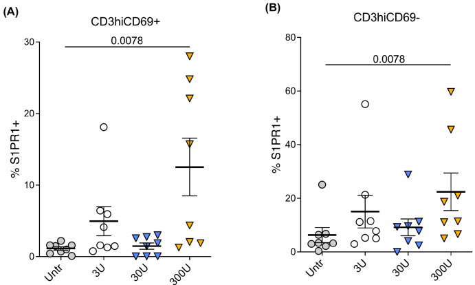
Figure 2.
Exogenous IFN-β increases S1PR1 protein expression in CD3hiCD69+ and CD3hiCD69− thymocytes in vitro. Thymocytes were cultured with different concentrations of IFN-β or media alone for 48 h (n = 8 individual thymus donors). S1PR1 expression in thymocyte subsets was measured by flow cytometry in eight different thymus tissues. (A) S1PR1 expression in CD3hiCD69+ thymocytes with different concentrations of IFN-β or media alone; (B) S1PR1 expression in mature CD3hiCD69− thymocytes with different concentrations of IFN-β or media alone. The mean with the standard error of the mean (SEM) is shown. Data were analyzed by one-way ANOVA using a Tukey HSD test for multiple comparisons. Overall, the data are statistically significant with p < 0.001. Tukey’s HSD Test for multiple comparisons found that the mean value of S1PR1 expression was significantly different between 300U and untreated (p = 0.0078 for both thymocyte populations).

