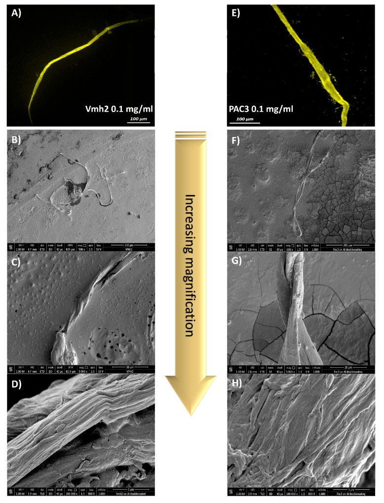
Figure 2.
Confocal laser microscope images of Vmh2 (A) and PAC3 (E) amyloid fibers stained by ThT. A total of 20 µL of protein solutions containing 3 µM ThT were deposited on a polystyrene support and air-dried prior to visualization. SEM images of Vmh2 (B–D) and PAC3 (F–H) fibers at different magnifications of 600×, 5000×, and 100,000×, respectively. A total of 20 µL of protein solutions were deposited on the surfaces, air-dried, and Au-Pd-coated prior to visualization.

