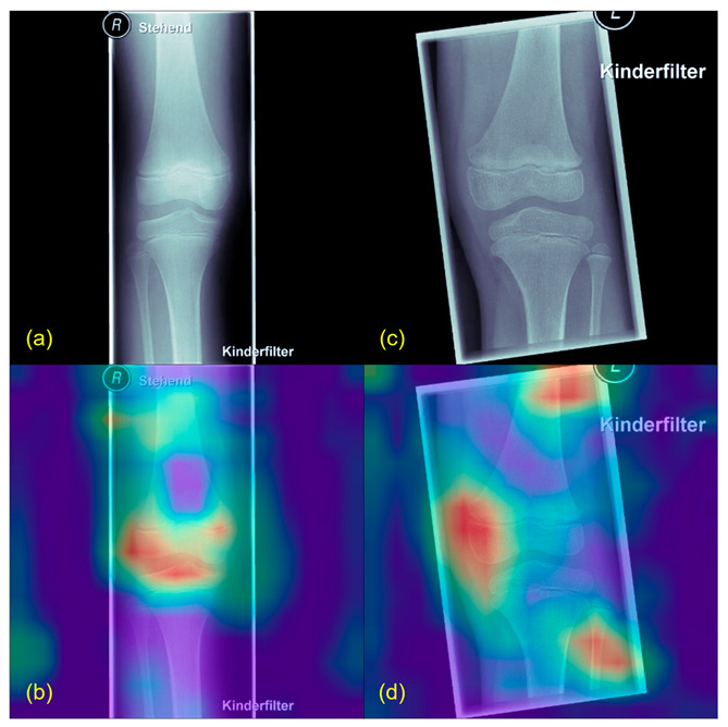
Figure 2.

Grad-CAM example from healthy patients. (a,c) show the original X-rays, (b,d) the Grad-CAMs of the same images. Highlighted in red are the parts used by the algorithm to find its conclusion. Patients classified as healthy usually showed a more diffuse Grad-CAM.
