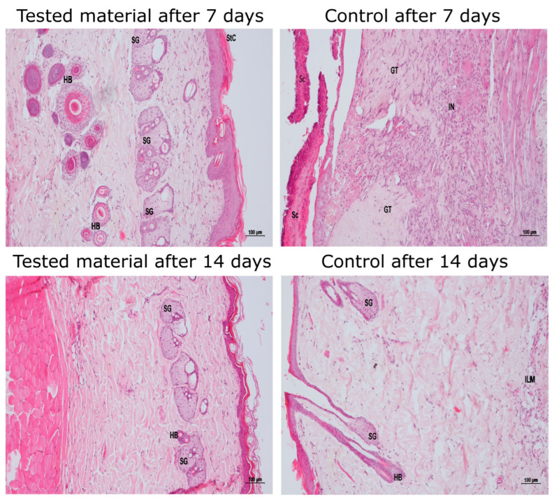
Figure 2.
Histopathological images of wounds. Visualized are two samples of the CMCS and the control hydrogels, collected from one rat on day 7 and from another rat on day 14; HB—hair bulb; SG—sebaceous gland; StC—stratum corneum; Sc—scab; GT—granulation tissue; I—inflammation; L—lymphocytic; M—monocytic; N—neutrophilic. A comprehensive analysis of all wounds is presented in Supplementary Materials Table S1.

