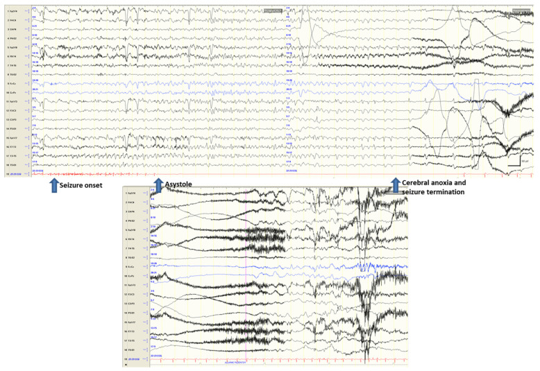
Figure 2.
Seizure onset from the left temporal region is followed (after about 8 s) by ictal asystole. During the 24 s of asystole, the EEG traces show the signs of hypoperfusion with progressive slowing of activity evolving into EEG suppression (with seizure termination after 19 s since asystole onset).

