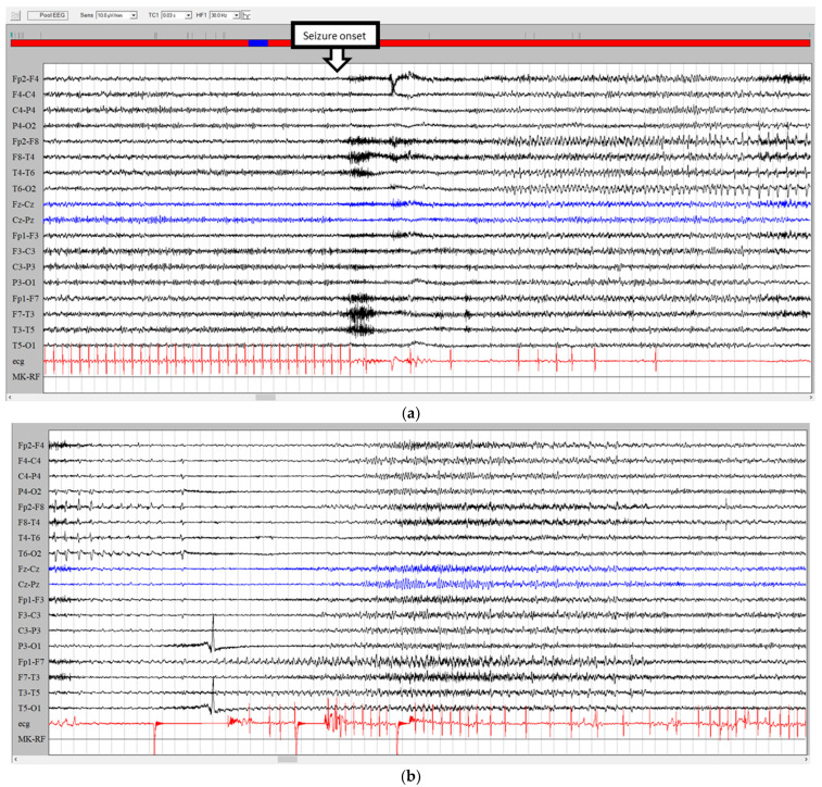
Figure 3.
(a) The EEG traces show a right frontotemporal recruiting activity rapidly associated with ictal arrhythmia (with a 28 s asystole). (b) The right ictal activity slows down as attended in the course of hypoxic attenuation of the tracing but unexpectedly restarts over the contralateral frontotemporal areas during asystole.

