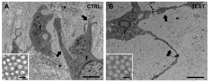
Figure 4.
Representative electron microscopy (EM) images of peri-implant soft tissues around the 2 abutments. The peri-implant soft tissue is primarily constituted by collagen fiber bundles and fibroblasts (f); (A) in CTRL samples, only a few collagen fibers assembled, forming small, scattered bundles, compared to TEST samples; (B) in TEST samples, collagen bundles were thick and dense, often covering a large area of the analyzed section. NOTE: In the analyzed area, collagen fibers are mainly “cross-sectioned” and appear as small circles (insets), assembling in bundles of different sizes. Asterisks in panel (A) refer to longitudinally oriented collagen fiber bundles. Black arrows point to fibroblast processes (f). Scale bars: 2 µm; insets, 0.1 µm.

