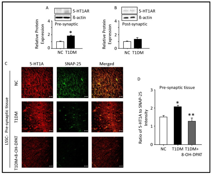
Figure 5.
Expression of 5-HT1AR in the presynaptic and postsynaptic fractions of the spinal cord lumbar region. Type 1 diabetes mellitus upregulates presynaptic (A) but not postsynaptic (B) spinal 5-HT1AR proteins. Immunofluorescence localization of presynaptic (C) 5-HT1AR in control, diabetic and diabetic administered with 8-OH-DPAT for 14 days. Quantitation of fluorescence intensity of 5-HT1AR relative to SNAP-25 (D). ImageLab 4.1 software was used to analyze protein bands of 5-HT1AR obtained with the ChemiDoc MP. Data are expressed as the mean ± SEM of values obtained from five animals/group. * Diabetic animals were significantly different from corresponding control values at p ≤ 0.05. ** 8-OH-DPAT-treated diabetic animals were significantly different from corresponding diabetic animals at p ≤ 0.05. Student’s t-test: one-way ANOVA followed by Dunnett’s post-test. Representative confocal images at magnification: 40X and scale bar: 20 µm. Abbreviations: NC—normal control, T1DM—type 1 diabetes mellitus, LSSC—lumbar segment of the spinal cord, SNAP-25—synaptosomal-associated protein-25, PSD-95—postsynaptic density-95, 5-HT1AR—5-HT1A receptors and 8-OH-DPAT—8-hydroxy-2-(dipropylamino)tetralin.

