Figure 2.

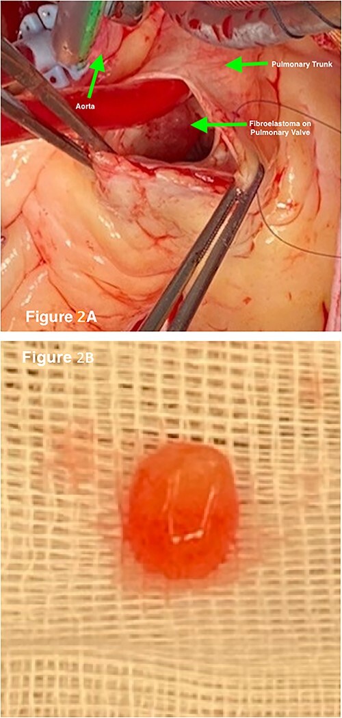
(a) Intraoperative imaging demonstrates the gelatinous lesion on the leaflet of the PV after opening the pulmonary trunk. (b) Excised PV papillary fibroelastoma.

(a) Intraoperative imaging demonstrates the gelatinous lesion on the leaflet of the PV after opening the pulmonary trunk. (b) Excised PV papillary fibroelastoma.