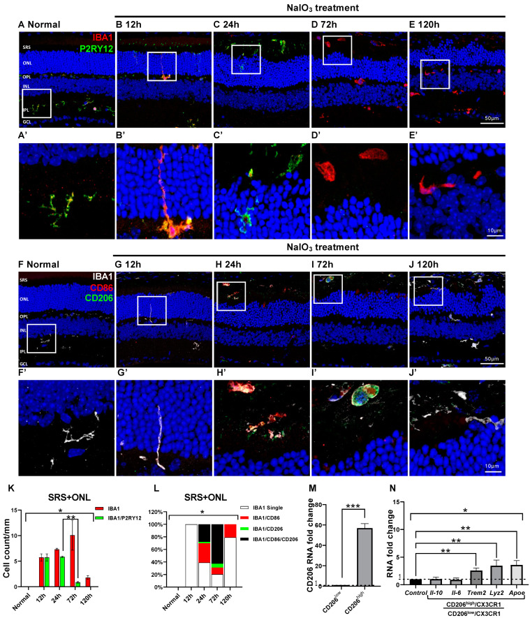
Figure 5.
Microglial cell response and characters in the NaIO3-induced RD. Anti-IBA1 antibody (red) and anti-P2RY12 antibody (green) were used to label the pan-microglial cells and innate microglial cells. (A–E). Representative images of IBA1/P2RY12-double-labeled vertical sections taken from normal (A) and RD retinas at 12 (B), 24 (C), 72 (D), and 120 h (E). Each boxed area is magnified in (A’–E’), respectively. Quantitative analyses of the IBA1/P2RY12-double-labeled microglial cells in the ONL and SRS showed significant loss of P2RY12 at 72 h (K). Representative images of IBA1/CD86/CD206-triple-labeled vertical sections taken from normal retina (F) and RD retinas at 12 (G), 24 (H), 72 (I), and 120 h (J). Each boxed area is magnified in (F’–J’), and the following three panels show IBA1-, CD86-, and CD206-channels, respectively. Scale bars: 50 μm (A–J), 10 μm (A’–J’). Quantitative analyses of the IBA1/CD86-double-labeled M1, IBA1/CD206-double-labeled M2, and IBA1/CD86/CD206-triple-labeled cells in the ONL and SRS (L). Moreover, 100% stacked column chart shows their proportions. The cell number was counted in the range of 700 μm from the optic disc in retinal vertical sections and presented as cell number per mm (n = 5). (M). The RNA expression level of the CD206 between CD206high/CX3CR1 and CD206low/CX3CR1 cells groups (n = 5). (N). The RNA expression level of the representative pro-inflammatory (Il-6), anti-inflammatory (Il-10), and phagocytosis-related (ApoE, Trem2, and Lyz2) genes between CD206high/CX3CR1 and CD206low/CX3CR1 microglial cells (n = 5). The fold-change of the RNA expression level was calculated using the delta-delta CT method after qRT-PCR. Data are presented as the mean ± S.E.M. * p < 0.05, one-way ANOVA with Tukey’s multiple comparison post-hoc test, ** p < 0.05, Tukey’s multiple comparison test, *** p < 0.05, Student’s t-test. GCL, ganglion cell layer; IPL, inner plexiform layer; INL, inner nuclear layer; OPL, outer plexiform layer; ONL, outer nuclear layer; SRS, subretinal space; Il-10, interleukin 10 gene; Il-6, interleukin 6 gene.

