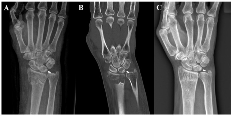
Figure 1.
Radiographs of a 70-year-old woman. (A) Preoperative posteroanterior radiograph and (B) coronal computed tomography image revealing sclerosis with cystic changes on the ulnar side of the lunate; (C) posteroanterior radiograph acquired after the removal of internal fixation at 12 months after surgery reveals the remnant sclerotic lesion with cystic change of the lunate.

