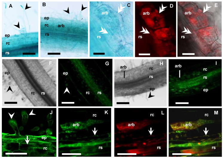
Figure 5.
MtAMF1;3 expressions in non-inoculated and mycorrhizal roots. (A–E) Medicago roots expressing MtAMF1;3pro:GUS (8 WAI, blue): (A) Non-inoculated root; (B–E) Mycorrhizal root; (C–E) Arbuscule-containing cell (80 µm vibrotome root sections); (C) Bright field view with GUS expression; (D,E) AM fungi inside cell, labelled with WGA-Texas Red antibody (excitation 555 nm, red channel). (F–M) Confocal microscope images of Medicago roots expressing UBQ3pro:GFP-MtAMF1;3 (8 WAI, green channel): (F) (DIC), (G,J) Non-inoculated root; (H) (DIC), (I,K–M) Mycorrhizal root; (F–I) Root overview, (J–M) Higher magnification of root cells; (J) GFP-MtAMF1;3 in root hairs and cortical cells of the Non-inoculated root; (K–M) GFP-MtAMF1;3 in arbuscule-containing cells of the mycorrhizal root: (K) GFP (green channel) view; (L) Red channel view—DsRED1 autofluorescence and WGA-TexasRed® antibody labelling of AM fungi cell walls; (M) Merged channels. On all images: ep = epidermis, rc = root cortex, rs = root stele, arb = arbuscule cells, black arrowhead shows root hairs, white double arrowhead indicates fungal hyphae, white arrow shows cell membranes. Scale bars: (A,B,F–I) 100 µm; (C–E,J–M) 20 µm.

