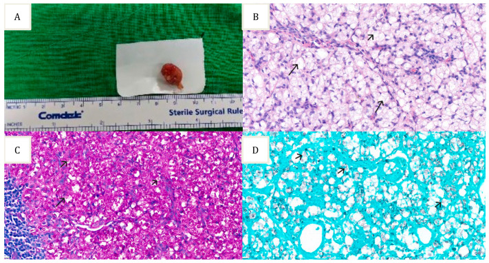
Figure 1.
Specimen of the inguinal lymph node: (A) Left inguinal lymph node excisional biopsy; (B) Hematoxylin and eosin stain showing lymph node tissue with numerous fungal elements; (C) Periodic-Acid Schiff stain highlighting the fungal elements; (D) Gomori’s Methenamine Silver stain highlight the fungal elements.

