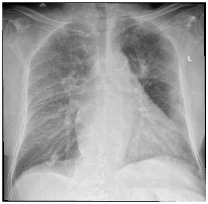
Figure 1.
Chest X-ray at admission—describing small left pleural effusion; bilateral diffuse reticule-micronodular interstitial lung pattern; questionable opacities at the level of the posterior left VI costal arch and right intercostal XI space; pulmonary vascular congestion; global enlarged heart; atherosclerotic aorta. L—left side.

