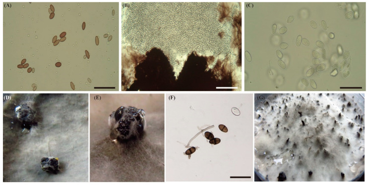
Figure 2.
Micrographs of conidial morphology and morphological culture structure on PDA. (A) Brown immature conidia of L. brasiliensis. Scale bar = 50 µm. (B) Pycnidia of L. hormozganensis were releasing immature conidia. Scale bar = 50 µm. (C) Immature conidia of L. hormozganensis. Scale bar = 50 µm. (D,E) Sporulation of L. hormozganensis and L. pseudotheobromae on PDA on the four-week colony. (F) Mature conidia of L. theobromae with dark-brown, one-septate with conspicuous vertical striations. Scale bar = 50 µm. (G) The appearance of pycnidia of L. theobromae on the surface of PDA with rich sporulation.

