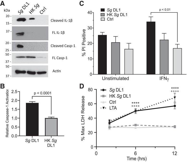
Fig. 4.
Live S. gordonii activates caspase-1 in macrophages. (A) Representative immunoblot showing caspase-1 and IL-1β cleavage. THP-1 cells were incubated with live or heat-killed (HK) S. gordonii DL1 for 24 h. Actin was used as a loading control. (B) Fold change in caspase-1 activation relative to THP-1 cells without bacteria added measured by YVAD-AFC fluorescence. (C) Propidium iodide (PI) acquisition by THP-1 cells after 24 h incubation. THP-1 cells were polarized with IFNγ or left unstimulated prior to incubation with live or HK S. gordonii. (D) LDH cytotoxicity after 2, 6, and 12 h incubation with live or HK S. gordonii with IFNγ-stimulated THP-1 cells. Transfected LTA was used as a positive control for pyroptosis. Percent release was determined compared with maximum LDH release by cells incubated with lysis buffer. Shown are mean ± SEM of 3 independent experiments. P value was calculated by unpaired t test (B) or ordinary 2-way analysis of variance followed by Dunnett's multiple comparisons in which each group was compared with control (+ indicates control vs LTA, * indicates control vs S. gordonii DL1) (C). Ctrl = control; Sg = Streptococcus gordonii.

