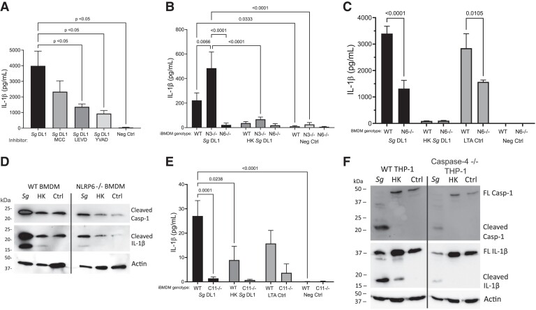
Fig. 5.
Determining inflammasome pathway activation by S. gordonii in macrophages. (A) IL-β release from PMA-differentiated, IFNγ-activated THP-1 macrophages with indicated inflammasome inhibitor added during incubation with S. gordonii for 6 h. MCC950 (MCC) indicates NLRP3 inhibitor, Ac-LEVD-CHO (LEVD) indicate scaspase-4/5 inhibitor, and Ac-YVAD-CHO (YVAD) indicates caspase-1 inhibitor. (B) IL-1β release as determined by enzyme-linked immunosorbent assay (ELISA) from IFNγ-stimulated immortalized BMDMs derived from WT, NLRP3 knockout, or NLRP6 knockout mice incubated with like or heat-killed (HK) S. gordonii for 24 h. (C) IL-1β release as determined by ELISA from BMDM derived from WT or NLRP6 knockout mice incubated with live or HK S. gordonii for 24 h. Transfected LTA (28 μg) was used as a positive control for NLRP6 activation.31 (D) Representative immunoblot showing caspase-1 and IL-1β cleavage. WT or NLRP6 knockout primary BMDMs were incubated with live or HK S. gordonii for 24 h. Actin was used as a loading control. (E) IL-1β release from caspase-11 knockout or WT iBMDMs incubated with live or HK S. gordonii for 24 h. (F) Representative immunoblot showing caspase-1 and IL-1β cleavage. WT or caspase-4 knockout THP-1 cells were incubated with live or HK S. gordonii for 6 h. Again, actin was used as a loading control. Shown are mean± SEM of 3 independent experiments. P values were calculated by 1-way analysis of variance followed by Dunnett's multiple comparisons (A, C, E) or 2-way analysis of variance followed by Sidak's multiple comparisons test (B). Ctrl = control; Sg = Streptococcus gordonii.

