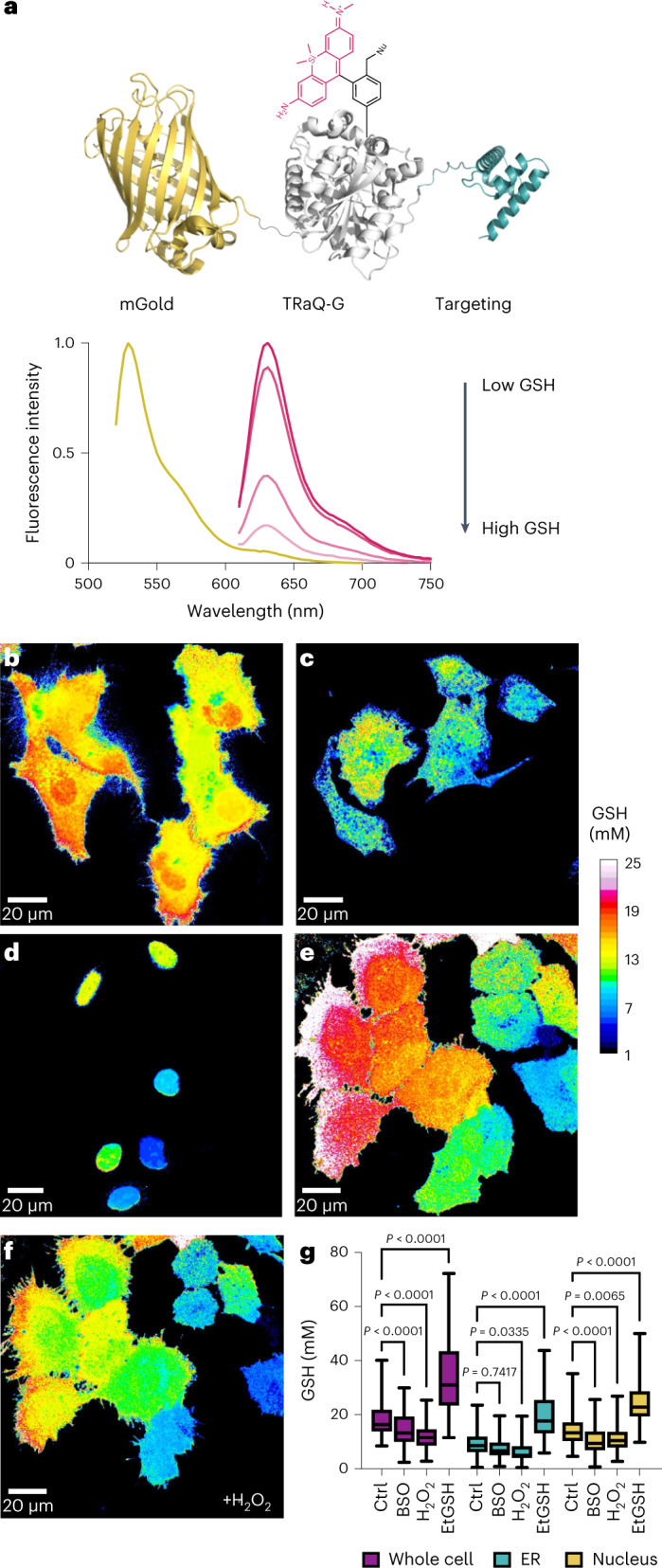
Fig. 3. GSH quantification in subcellular locations using TRaQ-G–mGold.

a, Schematic depiction of the GSH sensor TRaQ-G–mGold with a targeting peptide and mechanism of ratiometric sensing. b–d, Ratiometric imaging of GSH in whole cells (b), ER (c) and nuclei (d) using TRaQ-G–mGold. e,f, GSH quantification in cells before (e) and 20 min after (f) the addition of 1 mM H2O2. g, Quantification of GSH concentration in whole cells, the ER or untreated nuclei (ctrl) after the addition of 10 mM EtGSH (membrane-permeant GSH precursor), 1 mM BSO (inhibitor of GSH biosynthesis) or 1 mM H2O2 (oxidant). The TRaQ-G ligand concentration was 100 nM in all cases. Images were generated by dividing the intensity of mGold over that of TRaQ-G and relating this ratio to the calibration curve obtained with purified TRaQ-G–mGold. For display purposes, calibrated images were despeckled using Fiji (ImageJ). Statistical significance was evaluated by one-way analysis of variance (ANOVA) (Šídák’s multiple comparisons test) with N = 132, 142, 103, 127, 116, 129, 90, 118, 149, 176, 137 and 122 (from left to right) independent cells from three different passage numbers examined over three separate imaging sessions. Boxes represent 25th to 75th percentiles, the horizontal line represents the median, and whiskers extend from the minimum to the maximum.
