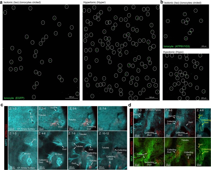
Extended Data Fig. 8. Osmotic stress in ALI cultures and a CF disease state in vivo induced ionocyte expansion.
a and b, Hyperosmotic (a) and hypoosmotic (b) ALI culture conditions induce ionocyte expansion. Ionocytes were localized either by lineage tracing using FOXI1-CreERT2 or ATP6V1G3 staining. Representative images of cultures derived from n = 5 donor ferrets for hyperosmotic stress and n = 4 ferrets for hypoosmotic stress. c and d, Additional panels related to Extended Data Fig. 7k. Whole mount trachea immunostaining demonstrating expanding ATP6V1G3+ ionocytes in KRT5+-enriched submucosal gland (SMG) ducts and NKA+ tubules of CFTRG551D/G551D ferrets (reared off VX-770). Confocal Z planes are indicated and show apical surface to SMG planes (c) or just the SMG plane (d). DO, duct opening. Representative images from n = 3 CF ferrets.

