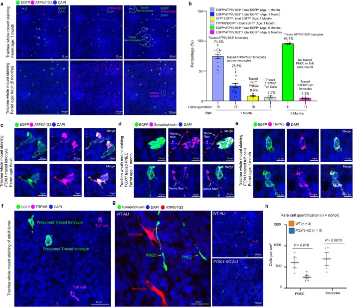
Extended Data Fig. 11. FOXI1-lineage rare cell progenitor has the capacity to generate ionocytes, PNECs, and tuft cells in vivo.
a, Confocal images of lineage-traced trachea following 5 sequential tamoxifen injections in FOXI1-CreERT2::ROSA-TG ferrets at 1 or 5 months of age (harvested one week after the last tamoxifen injection). Traced EGFP+ATP6V1G3+ ionocytes and EGFP+ATP6V1G3– non-ionocyte were identified in one month old trachea (upper panel). In adult FOXI1-CreERT2::ROSA-TG ferret trachea, only traced EGFP+ATP6V1G3+ ionocytes were observed (lower panel). Representative images from n = 3 independent ferrets were traced in each group. b, Percentage of FOXI1-CreERT2 labeled EGFP+ cells in the trachea for the indicated phenotypic classifications following colocalization with markers for ionocytes (ATP6V1G3), PNEC (synaptophysin/SYP), and tuft cells (TRPM5) at 1 and 5 months of age (Mean ± s.e.m.; n = 3 ferrets quantified for each age; each datapoint represents quantification from a 1.41 mm2 area for the number of fields indicated). Lineage tracing of 1 month old FOXI1-CreERT2::ROSA-TG ferrets demonstrated FOXI1-lineage cells (EGFP+) were composed of ionocytes (74.5%), PNEC (8.6%) and tuft cells (6.5%). Lineage tracing of 5 month old (adult) FOXI1-CreERT2::ROSA-TG ferrets demonstrated 95.7% of FOXI1-lineage cells (EGFP+) were ATP6V1G3+ ionocytes. The 4.3% EGFP+ATP6V1G3– cells in adult animals is consistent with a small fraction of mature ionocytes (~4%) expressing no ATP6V1G3 by scRNAseq. c, Maximum intensity projection of traced EGFP+ATPV61G3+ pulmonary ionocytes from adult FOXI1-CreERT2::ROSA-TG ferret trachea. Representative image from n = 3 ferrets. d, Maximum intensity projection of traced EGFP+SYP+ PNECs from one month old FOXI1-CreERT2::ROSA-TG ferret trachea. Images show sensory nerves associated with PNECs. Representative image from n = 3 ferrets. e, Maximum intensity projection of traced EGFP+TRPM5+ tuft cells from one month old FOXI1-CreERT2::ROSA-TG ferret trachea. Representative image from n = 3 ferrets. f, Representative image of adult FOXI1-CreERT2::ROSA-TG ferret trachea following lineage tracing demonstrating the lack of TRPM5 staining in EGFP+ presumed ionocytes. Representative image from n = 3 ferrets. g, Representative confocal image of ionocytes (ATP6V1G3+) and PNECs (SYP+) in epithelia from WT and FOXI1-KO air-liquid interface (ALI) cultures. n = 4 ferrets (WT), n = 5 ferrets (FOXI1-KO). h, Frequency of ionocytes (ATP6V1G3+) and PNECs (SYP+) in epithelia from FOXI1-KO and WT ALI cultures (n = ferret donors, 3-5 independent cultures quantified from each donor and averaged). Graphs show the mean ± s.e.m. Statistical significance was determined by two-tailed Student’s t-test.

