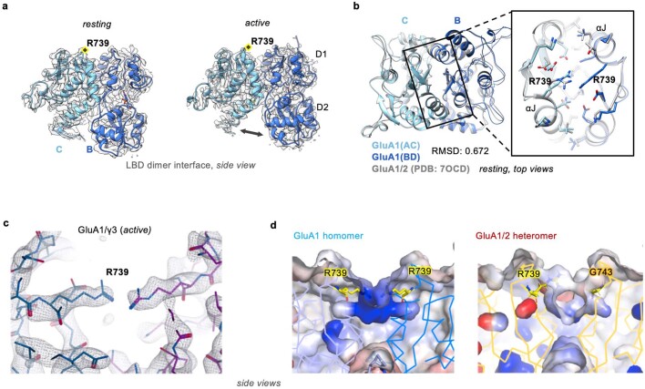
Extended Data Fig. 7. Features of resting and active GluA1/γ3 LBD dimers.
a, Side view of cryo-EM density and model of GluA1/γ3 LBD dimer in resting and active states. b, Top of resting GluA1/γ3 LBD dimer interfaces aligned to those from a GluA1/2 heteromer (PDB: 7OCD), showing close similarity despite the presence of the R739 bridge. c, A zoomed view into the arginine bridge between R739 residues resulting from lack of an R/G editing site in active GluA1 supported by strong density shown for the open state as seen for R/G-unedited GluA2 LBD (PDB: 2UXA). A similar arrangement is also seen in the resting state (not shown). d, Electrostatic maps from APBS showing the dispersal of positive charge around R739 in GluA1 compared to the GluA1/2 heteromer, contoured from −10kT/e (red) to + 10kT/e (blue).

