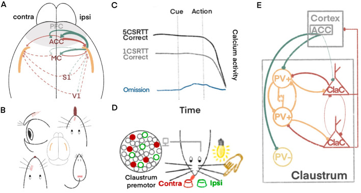
Fig. 1.
Motor control mechanisms of the claustrum. A The claustrum mainly receives contralateral projections from the PFC, ACC, and MC, with the strongest projections coming from the ACC. The output connections from the claustrum to the cortex tend to have ipsilateral dominance. B The claustrum regulates movements of sensory organs such as eyelids, the tongue, whiskers, and the tail. C The overall activation of the nucleus is higher when performing complex motor tasks than when performing simple motor tasks, and its overall neuronal activity runs through the task prompt until the end of the action. D Electrodes are implanted on the side of the claustrum and monitored, with two licking points located on the ipsilateral and contralateral sides of the implanted electrodes. Red indicates claustrocortical (ClaC) neurons with contralateral preference and green indicates neurons with ipsilateral preference. Red neurons are labeled as solid, indicating that ClaC neurons with contralateral preference are synchronously activated before the mouse licks the contralateral side. E The claustrum predominantly receives modulation from the ACC during motor control, which is tuned by synchronization of internal microcircuits followed by signal output to various parts of the cortex. Three types of claustrum neurons, aspiny PV+ interneurons, aspiny PV− interneurons, and ClaC are distinguished. The same color is used for homogenous neurons and synaptic markers. Extruded lines at the ends indicate chemical synapses, parallel lines close to each other indicate gap junction connections and thin lines indicate weak connections.

