Figure 7.
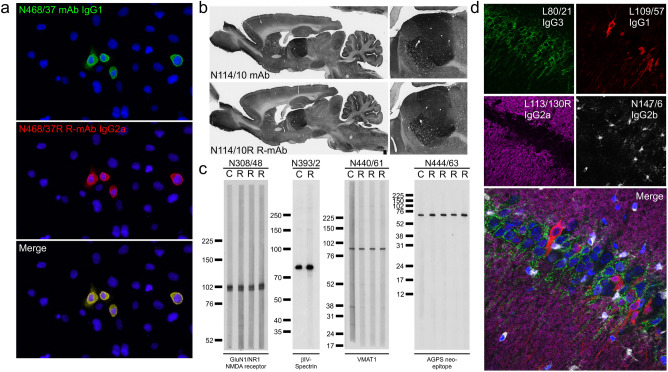
R-mAb evaluation. (a) Immunofluorescence immunocytochemistry on transiently transfected COS-1 cells. Cells were transfected with a plasmid encoding mouse THIK-2 and double immunolabeled with the progenitor N468/37 IgG1 mAb (green) and the N468/37R IgG2a R-mAb (red). Hoechst nuclear labelling is shown in blue. (b) Immunoperoxidase/DAB immunohistochemistry on rat brain sections comparing immunolabeling with the anti-HCN4 hybridoma-generated N114/10 mAb to the N114/10R R-mAb. (c) Strip immunoblots on rat brain samples showing side-by-side comparisons of hybridoma-generated conventional progenitor mAb samples “C” and transfected cell generated R-mAb samples “R”. Numbers to the left of each set denote mobility of molecular weight standards in kD. (d) Immunofluorescence immunohistochemistry on rat brain sections. Multiplex immunolabeling with four mouse mAbs including the subclass-switched R-mAb L113/130R in adult rat hippocampal CA1.

