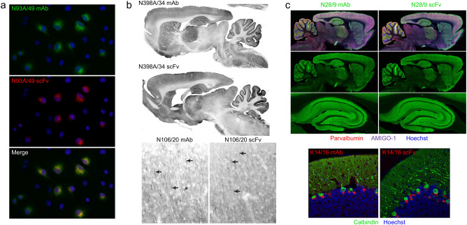
Figure 8.
scFv evaluation. (a) Immunofluorescence immunocytochemistry on transiently transfected COS-1 cells. Cells were transfected with a plasmid encoding the mouse GABAB R1 receptor and double immunolabeled with the progenitor N93A/49 mouse mAb (green) and the N93A/49 scFv (red). Hoechst nuclear labelling is shown in blue. (b) Immunoperoxidase/DAB immunohistochemistry on rat brain sagittal sections. The top pair of images show immunolabelling with the anti-GABAA receptor α4 hybridoma-generated N398A/34 mAb (top section) and the N398A/34 scFv (bottom section). The bottom pair of images show immunolabelling in neocortex with the anti-Ankyrin-G hybridoma-generated N106/20 mAb (left section) and the N106/20 scFv (right section). (c) Immunofluorescence immunohistochemistry on rat brain sagittal sections. The top set of images show multiplex immunolabeling of brain (top two rows) and hippocampus (bottom row) with the anti-VGlut1 hybridoma-generated N28/9 mAb (IgG1) (left section) and the N28/9 scFv (right section) in green. The sections were also labelled with the mouse mAbs anti-Parvalbumin L114/3 mAb (IgG2a) in red and the anti-Amigo-1 L86A/37 mAb (IgG2b) in purple. Hoechst nuclear labelling is shown in blue. The bottom pair of images show multiplex immunolabeling of rat cerebellum with the anti-Kv1.2K+ channel hybridoma-generated K14/16 mAb (IgG2b) (left section) and the K14/16 scFv (right section) in green. The sections were also labelled with the mouse anti-Calbindin L109/39 mAb (IgG2b). Hoechst nuclear labelling is shown in blue.

