Fig. 1.
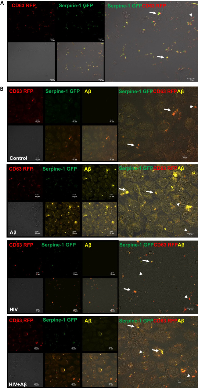
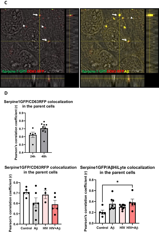
Serpine-1 is concentrated in vesicular structures in brain endothelial cells. HBMEC were transfected with the Serpine-1 GFP and CD63 RFP plasmids (A) and 24 h after transfection, cells were exposed to HIV-1 (30 ng p24/ml) and/or 100 nM Aβ (1–40) HiLyte 647 for 48 h (B). A The images represent live imaging of Serpine-1 GFP and CD63 RFP (arrow heads) in the parent HBMEC 24 h after transfection. Scale bar: 10 μm. B Serpine-1 GFP, CD63 RFP, or Aβ HiLyte 647-positive fluorescence (arrow heads) in the fixed parent cells 48 h after vehicle (control), HIV-1, and/or Aβ exposure. Arrows indicate overlapping positive fluorescence of Serpine-1 GFP, CD63 RFP, and/or Aβ HiLyte 647. Scale bar: 10 μm. Representative images from four experiments. C Confocal z-stack imaging of Serpine-1 GFP, CD63 RFP, and Aβ HiLyte 647 colocalization (arrows) in the fixed parent cells 48 h after HIV-1 and Aβ exposure. Scale bar: 5 μm. D Quantification of colocalization of Serpine-1 GFP and CD63 RFP in the live parent cells after transfection (upper graph), unpaired t-test; Colocalization of Serpine-1 GFP and CD63 RFP (lower left graph) and Serpine-1 GFP and Aβ HiLyte 647 (lower right graph) in the fixed parent cells 48 h after HIV-1 and/or Aβ exposure. Values are mean ± SEM, n = 3–9. One- and two-way ANOVA with Tukey’s multiple comparisons test. *Statistically significant at p < 0.05


