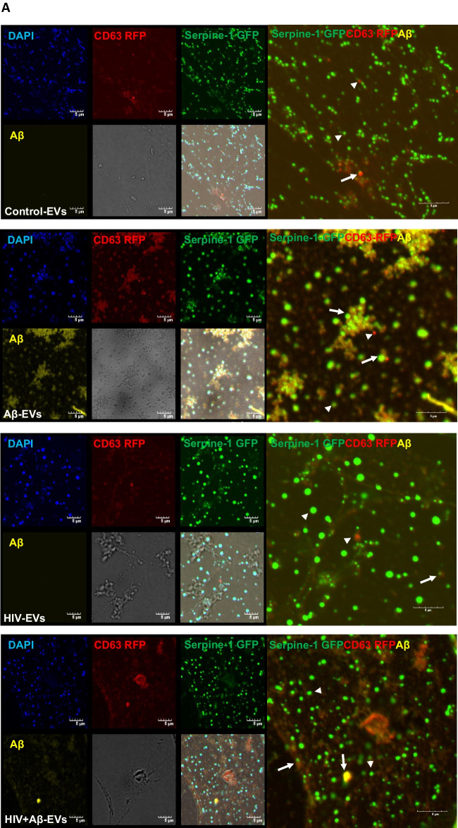
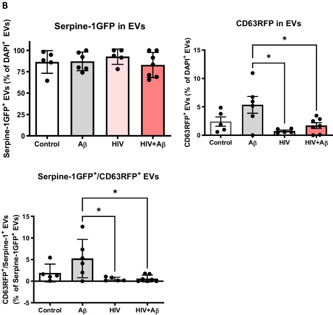
Fig. 2.
Serpine-1 is released in EVs from control, Aβ, and/or HIV-1 exposed brain endothelial cells (A) Visualization by confocal microscopy of Serpine-1 GFP (green), CD63 RFP (red), and Aβ (1–40) HiLyte (yellow) (arrow heads) in EVs isolated from media of transfected and treated HBMEC as in Fig. 1. Examples of overlapping fluorescence of Serpine-1 GFP, CD63 RFP, and/or Aβ (1–40) HiLyte associated with EVs are indicated by arrows. DAPI stains the genetic material in EVs. Representative images from three experiments. Scale bar: 5 μm. B Quantification of Serpine-1 GFP-positive, CD63 RFP-positive, and Serpine-1 GFP/CD63 RFP-double positive EVs. Values are mean ± SEM, n = 5–7. One- and two-way ANOVA with Šídák's and Tukey’s multiple comparisons tests. *Statistically significant at p < 0.05


