FIGURE 2.

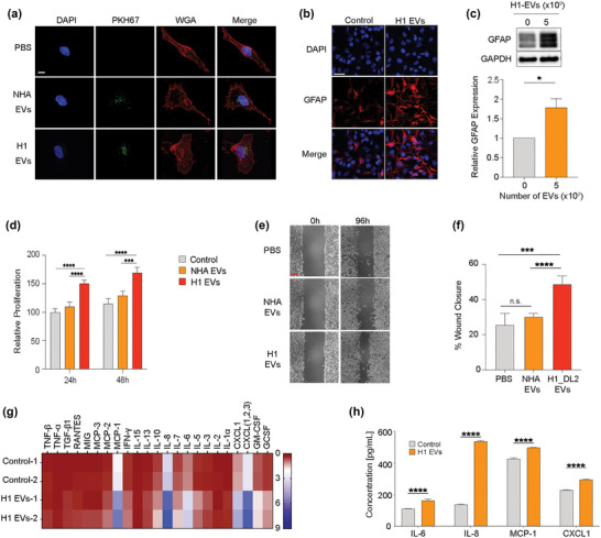
MBM‐derived EVs induce activation in NHA. (a) NHA cultured with 5.0 × 10⁹ EVs of PKH67‐stained (green) NHA‐ or H1‐EVs for 48 h to show uptake. Nuclei stained with DAPI (blue), membrane stained with WGA‐Texas Red (red). Magnification (100X). Scale bar = 10 μm. (b) Representative images of ICC‐stained NHA cells cultured with H1‐EVs or PBS control for 48 h. Merged images of DAPI (blue) and GFAP (red). Magnification 20X. Scale bar = 20 μm. (c) Western blot analysis of GFAP in NHA cells after culturing with 5.0 × 10⁹ H1‐EVs or PBS for 48 h and subsequent quantification. (d) CCK8 proliferation assay of NHA cells cultured with 5.0 × 10⁹ NHA‐ or H1‐EVs over 48 h. (e) Representative micrographs at the start and completion of a 96 h NHA wound healing assay co‐cultured with 5.0 × 10⁹ NHA‐ or H1‐EVs. Scale bar: 300 μm. (f) Quantification of wound healing assay as percentage wound closure after 96 h. (g) Heatmap of a cytokine array of NHA conditioned media after culturing with 5.0 × 10⁹ H1‐EVs or PBS for 48 h. (h) ELISA validation performed on NHA conditioned media after co‐culture with 5.0 × 10⁹ H1‐EVs. ELISA of top four upregulated cytokines from cytokine array, IL‐6, IL‐8, MCP‐1 and CXCL1, were used. n.s.= not significant *p < 0.05, ***p < 0.001, ****p < 0.0001.
