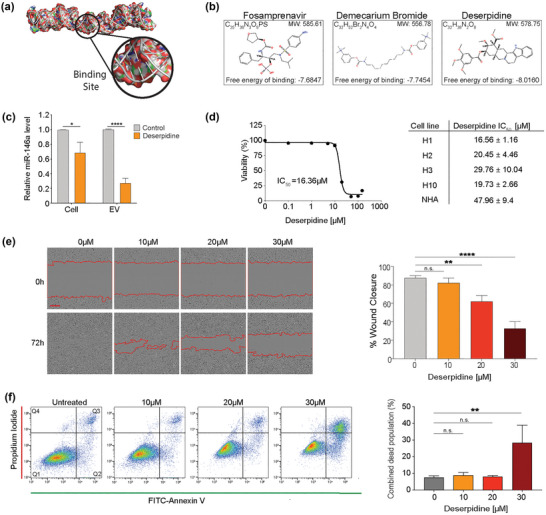
FIGURE 7.

Deserpidine reduces the expression of miR‐146a‐5p in vitro and reduces the viability and proliferation of MBM cells. (a) Schematic representation of the 3D structure of the miR‐146a‐5p binding site for high‐throughput molecular docking analysis. (b) Schematic representation of the top 3 binding partners of miR‐146a‐5p available for purchase and predicted to cross the blood brain barrier. (c) qPCR analysis of miR‐146a‐5p expression in H1 cells and EVs after deserpidine treatment. (d) Representative IC50 survival curve of H1 cells after treatment with increasing drug concentrations (0.01–150 μM) for 72 h and corresponding deserpidine IC50 doses of all MBM cell lines and NHA. (e) Representative images from wound healing assay of H1 cells after treatment with various concentrations of deserpidine and quantification of % wound closure at 72 h. Scale bar = 300 μm. (f) Annexin V‐FITC and PI staining to assess apoptosis by flow cytometry in H1 cells treated with increasing concentrations of deserpidine for 72 h and corresponding quantification of live cell and apoptotic cell populations (Q2 + Q3) represented as percentage of total cell population. All data displayed from three independent experiments. Quadrant 1: Live cells. Quadrant 2: Early apoptosis. Quadrant 3: Late apoptosis. Quadrant 4: Necrosis. n.s.= not significant, *p < 0.05, **p < 0.01, ****p < 0.0001. N = 3.
