Figure 5.
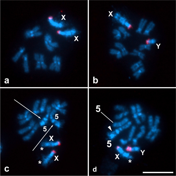
Localization of rDNA clusters in mitotic chromosomes of Ceratitis capitata by FISH with the 18 rDNA probe (red signals). Chromosomes, obtained from the brains of wild-type larvae (a, b) and T(X;5) larvae (c, d), were counterstained with DAPI (light blue). X and Y symbols denote sex chromosomes, symbol 5 stands for chromosome 5. Bar = 10 µm. (a) Female mitotic metaphase with two X chromosomes, each with a terminal rDNA cluster, and five pairs of autosomes. (b) Male mitotic metaphase with X and Y chromosomes, each with a terminal rDNA cluster, and five pairs of autosomes. (c) Female mitotic metaphase showing reciprocal translocations between two X chromosomes (asterisks), each with a terminal cluster of rDNA, and two chromosomes 5 (arrows). (d) Male mitotic prometaphase showing a T(X;5) translocation on the X chromosome (asterisk) and a T(5;X) translocation on one chromosome 5 (arrowhead); both the X and Y chromosomes show terminal clusters of rDNA (note that the rDNA clusters on the Y chromosome appear duplicated).

