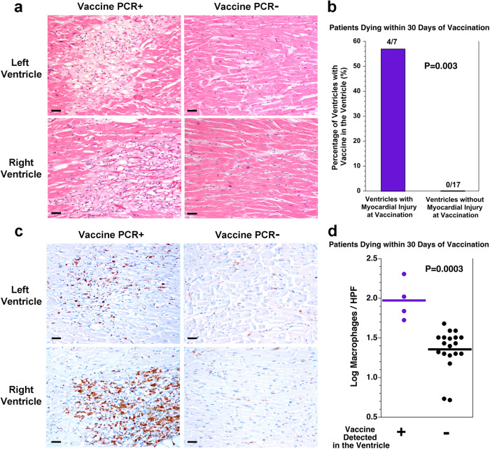
Fig. 3. SARS-CoV-2 vaccine in the heart is associated with healing myocardial injury at the time of vaccination and macrophage infiltration in the myocardium.
a Depicted are histologic images of the myocardium from both the left and right ventricles of the heart with either vaccine detected in the sample or not detected in the sample. For the two samples in which vaccine was detected, there was healing myocardial injury (left), compared with no healing myocardial injury in the two samples in which vaccine was not detected (right). Scale bars indicate 40 microns. b For the 12 patients dying within 30 days of vaccination, myocardial injury was present at the time of vaccination in 4 of the 7 ventricular samples with vaccine in the heart but none of the 17 ventricular samples without vaccine in the heart (P = 0.003, Fisher exact test). c Depicted are immunohistochemical stains for the macrophage marker CD68 on the myocardium from both the left and right ventricles of the heart with either vaccine detected in the sample or not detected in the sample. For the two samples in which vaccine was detected, there are more macrophages (left, brown stain), compared with the two samples in which vaccine was not detected (right). Scale bars indicate 40 microns. d Quantitation of the macrophages revealed more macrophages per 400× high power field (HPF) in the ventricular samples in which vaccine was detected than in the ventricular samples in which vaccine was not detected (P = 0.0003, t test, n = 4 vs n = 19, t = 4.3782, df = 21, difference between means = 0.62, 95% CI: 0.21–0.91). Tissue from one ventricle without vaccine detection and without myocyte injury was not available for CD68 immunohistochemistry.

