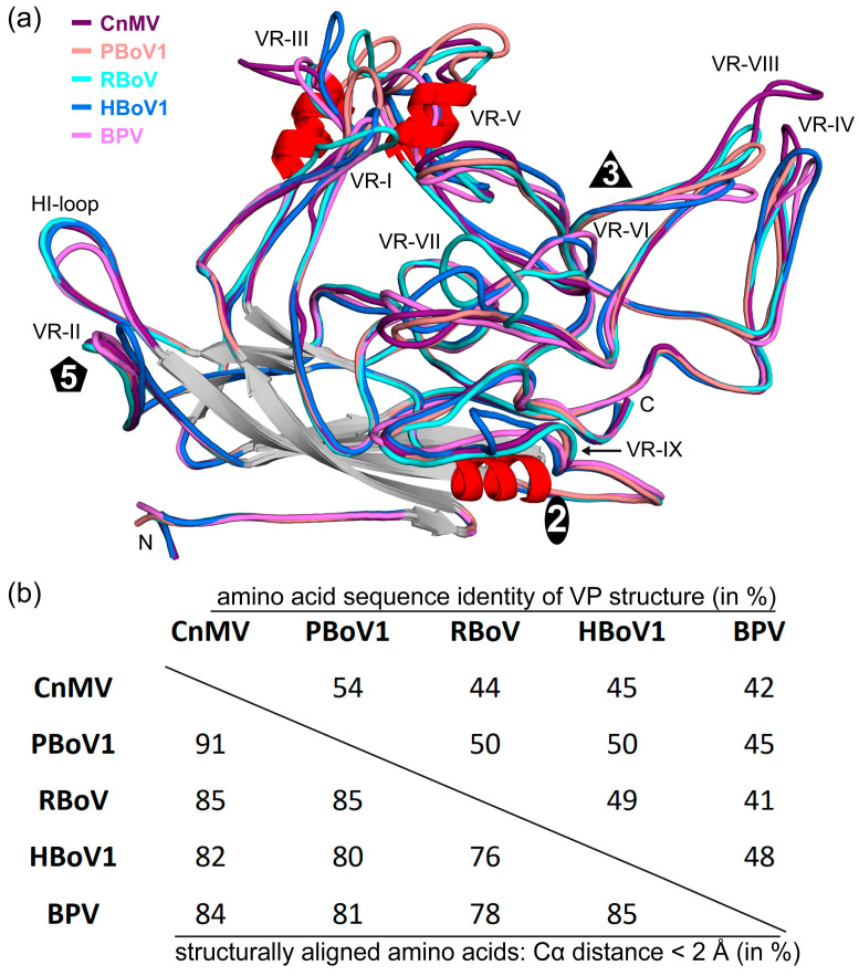
Figure 4.
Sequence and structure comparison within the Bocaparvovirus genus. (a) Superposition of CnMV (purple), PBoV1 (salmon), RBoV (cyan), HBoV1 (blue), and BPV (pink). The variable regions, the N- and C-termini, and the approximate location of the icosahedral two-, three-, and fivefold axes are shown. (b) Amino acid sequence identity comparison of the structurally ordered VP region for the given viruses (top right, in %). The structural similarity is shown in the bottom left corner and was defined as the percentage of aligned Cα atoms of the amino acid chain within 2 Å distance when the capsid structures were superposed.

