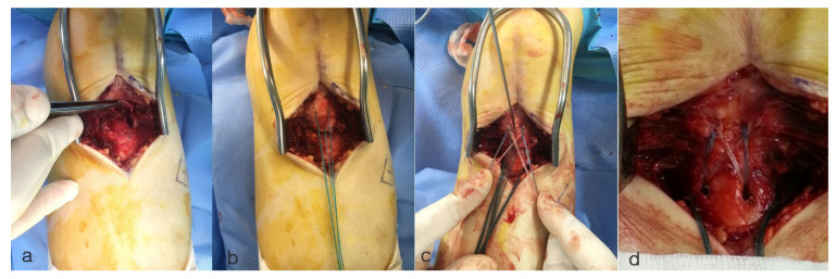
Figure 2.
Intra-operative images. (a) Image depicts a tibial tuberosity avulsion fracture, as indicated by the forceps holding the avulsed fragment. (b) The avulsed tendon was grasped and pulled using No. 2 Ethibond suture, and an appropriate suture anchor location was identified. (c) Image shows the fixation method using the suture bridge technique. A single suture limb from each anchor was secured over the tibial tuberosity and fastened to the distally positioned footprint anchors. (d) A well-reduced state was observed with the suture bridge repair technique.

