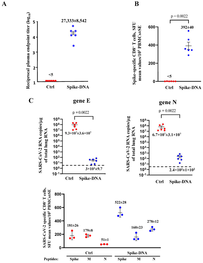
Figure 2.
Spike-specific immune responses and viral replication in spike-DNA injected mice. (A) Detection of anti-S1 antibodies in plasma of K18-hACE2 mice i.m. injected with either void (Ctrl, 6 mice) or spike-expressing (6 mice) DNA vectors. The log10 of reciprocal endpoint titers is shown, together with intragroup mean ± SE. (B) Detection of SARS-CoV-2-spike-specific CD8+ T-cells in PBMCs. A total of 105 PBMCs were incubated overnight with 5 μg/mL of either unrelated or spike-specific peptide in IFN-γ ELISpot microwells. The numbers are shown of spot-forming units (SFUs)/well calculated as mean values of triplicates after subtraction of the mean spot numbers detected in wells of PBMCs treated with an unspecific peptide (<5 spots for each cell culture tested). The intragroup mean values ±SE are reported. (C) Viral loads in the lungs of immunized/infected mice. Four and six days after the challenge, injected mice (6 per group) were sacrificed, and the lungs were processed for the extraction of total RNA. Then, 1 μg of total RNA from each infected mouse was analyzed by RT-qPCR for the presence of both SARS-CoV-2 E- and N-specific RNAs. The numbers are shown of both E and N viral RNA copies, amplified from total RNA isolated from the lungs of each animal, together with intragroup mean values ± SE. Dotted lines signal the detection limits of the assay. (D) Detection of spike-, M-, and N-specific CD8+ T-cells in PBMCs isolated 6 days after infection. A total of 105 PBMCs were incubated overnight with or without 5 μg/mL of either unrelated spike-, M-, or N-specific peptides in IFN-γ ELISpot microwells. The numbers of SFUs/well calculated are shown as mean values of triplicates after subtraction of the mean SFUs detected in wells of PBMCs treated with unspecific peptides (<4 spots for each cell culture tested). The intragroup mean values ±SE are reported.

