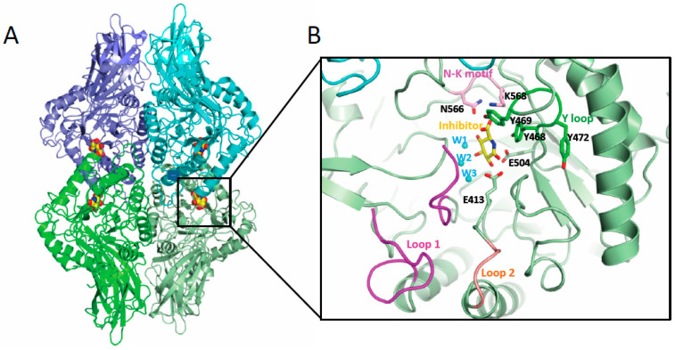
Figure 2.
Gut microbial GUS structure. (A) Quaternary architecture of GH2 family GUS. The PDB 3K4D corresponding to EcGUS bound to the glucaro-d-lactam inhibitor is shown. Each dimer is represented in different shades of green and blue and the inhibitor is shown in sphere representation (yellow, red and blue represent carbon, oxygen and nitrogen atoms). (B) Detail of the active center of one of the subunits showing some structural elements involved in conjugated E binding in GUS enzymes including Loop 1 (magenta), Loop 2 (salmon), N-K motif (pink) and Y loop (dark green). Also, those residues and water molecules (in cyan) involved in substrate binding and catalysis are labelled.

